A multifaceted screening procedure of high accuracy and detection range.
The transvaginal ultrasound (TVUS) is a crucial diagnostic tool for detecting mild and severe gynecological conditions or evaluating infertility and early pregnancy.
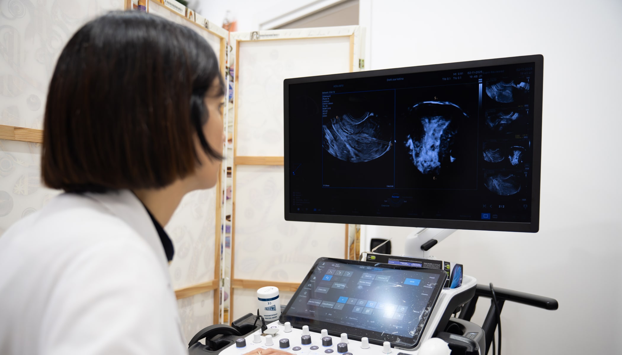
A TVUS is conducted from inside the vagina, offering comparatively higher resolution images of the female reproductive organs and surrounding structures. Its use of high-frequency sound waves and its increased proximity to the examined areas make it more accurate for detecting abnormalities (such as fibroids, polyps, tumors, cysts, ectopic pregnancies) and for returning vital measurements (such as endometrial thickness, follicle count, tubal dilation, cervical length).
The examination is completely safe for pregnancy (no radiation), but it’s avoided in virgins due to a high risk of hymen injury.
Preparation for a transvaginal ultrasound requires a normal hygiene routine and an empty bladder for clearer images. The procedure can take place at any phase of the menstrual cycle. It typically lasts 15-30 minutes, and it’s usually painless, but may cause mild discomfort similar to a pelvic exam:
A sterile, gel-covered probe is inserted into the vagina
The operator moves the probe to capture images from different angles
If you have any questions before the examination, don’t hesitate to ask your specialist.
The TVUS results are typically available within a few days and are sent to your OB-GYN for evaluation of severity and possible follow-up exams.
Key areas covered include the uterus, cervix, ovaries, and adnexa. Abnormal findings may range from minor or mild, which may need monitoring, to highly concerning, which may require further investigation or even emergency intervention.
The transvaginal ultrasound is one of the main sonogram types used to monitor early pregnancy and fetal health.
For appointments, call my office every Thursday between 9.30 and 15.30.


